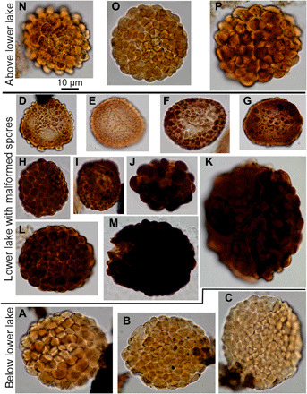
Fig. 4. V. nitidus spores from below, in, and above the D-C boundary showing UV-B malformations.

(A) to (C) are from below the boundary (Stensiö Bjerg) and have the characteristic packed verrucae sculpture of V. nitidus. (D) to (K) are from the lower lake (Rebild Bakker), with (H) to (M) more strongly pigmented and more regular sculpture with a wider range of diameter than normal. (D) to (G) are paler in color and smaller in size and have irregularly developed sculpture. (N) to (P) are normal specimens of V. nitidus from the upper lake bed (Rebild Bakker). Sample and slide numbers plus England Finder coordinates are in table S4.
