. 2020 Apr 20;9:e53479. doi: 10.7554/eLife.53479

Figure 6. Conformational change at Helices G-I and repositioning of Glnin (Helix H).

(A) View of one CLC subunit, highlighting Repeat 1 in pink and Repeat 2 in teal. Helix R (not part of the repeats) is shown in grey. (B) Movement of Helices G-I (part of Repeat 1) relative to Helices K, M, N (Repeat 2). The compared helices are shown in ribbon, with QQQ in purple and WT in gray (Helices K, M, N) or yellow (G–I). Other helices (WT) are shown as transparent cylinders. The inset illustrates how the movement of Helix H away from Helix N creates space to avoid steric conflict with Glnex in the ‘out’ position. (C) Movement of Helices G-I relative to other helices in Repeat 1. The compared helices are shown in ribbon, with QQQ in purple and WT in gray (Helices C-E) or yellow (G–I). Other helices (WT) are shown as transparent cylinders. (D) Close-up view showing movement of Helix H (containing Glnin) away from Helix D. (E) Conformational change at Glnin moves it away from Q113. (F) Movement of Glnin to the hydrophobic core of the protein brings it to within 6 Å of Glnex. Electron density for Glnex, Glnin, and an intervening water molecule is shown in mesh.

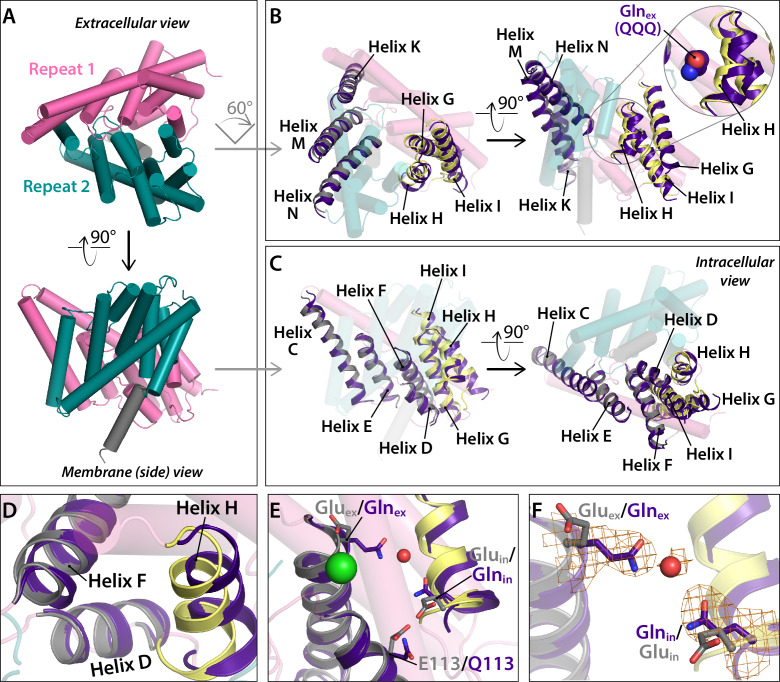
Figure 6.

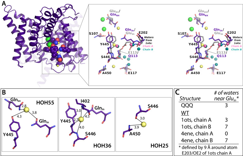
Figure 6—figure supplement 1. Crystallographic water molecules near Glnin.

Figure 6—figure supplement 1.

(A) Left: 1 subunit of QQQ CLC-ec1. The three water molecules within 9 Å of Glnin (see panel C) are shown as yellow spheres. Glnex, Glnin, Cl ions, and inner-gate residues Y445 and S107 are shown in spacefill. Right: zoomed in stereoview of the boxed region, showing QQQ (purple) overlaid with WT (1ots) CLC-ec1 (grey). Water molecules seen in subunits A and B of 1ots are shown in pink (three water molecules) and teal (seven water molecules) respectively. (B) Close-up views of the three crystallographic water molecules near Glnin in QQQ, showing distances of potential coordinating atoms. (C) Comparison of water molecules near Glnin/Gluin in QQQ, 1ots, and 4ene, a CLC-ec1 construct with trimmed N- and C-termini, which behaves functionally like WT (Lim et al., 2012). 1ots and 4ene have the highest resolution among the reported structures with WT sequences. Overall, the total number of modeled waters associated with the protein dimer in each structure is 110 (QQQ), 167 (1ots), and 52 (4ene). The higher number of water molecules in 1ots is due to the modeling of waters with less stringent parameters (more clashes of waters with protein atoms and the modeling of waters farther than 3.5 Å from the protein atoms). 1ots also has high R factors (Rwork = 0.26 and Rfree = 0.30).