
Figure 6.
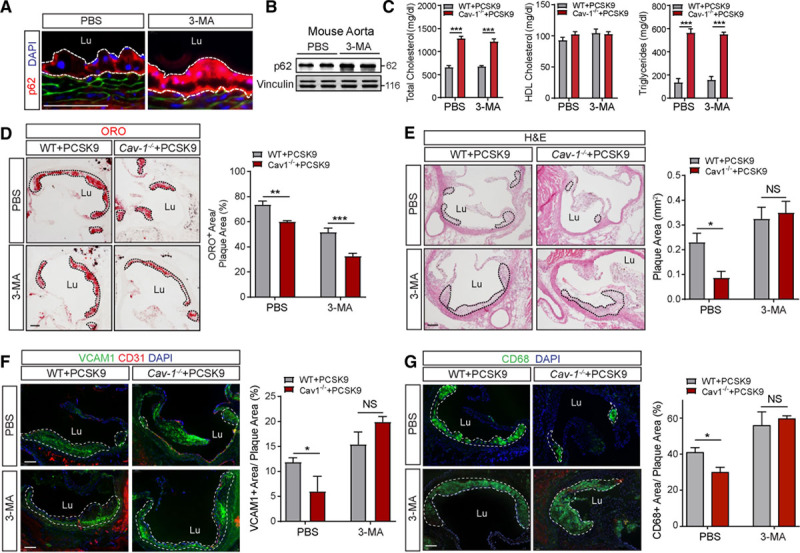
Inhibition of autophagy attenuates the atheroprotection observed in the absence of Cav-1 (caveolin-1). A, Representative immunofluorescence images of p62 staining in the lesser aortic curvature of WT mice injected with adeno-associated virus (AAV)-PCSK9, treated with 3-methyladenine (3-MA) or PBS, and fed a Western-type diet (WD) for 4 wk. B, Western blot analysis of p62 expression in aortas from WT (wild type) mice treated as indicated in A. Vinculin was used as loading control. C, Total cholesterol, HDL (high-density lipoprotein) cholesterol, and triglyceride levels in WT and Cav-1−/− mice injected with AAV-PCSK9, treated with 3-MA, and fed a WD for 4 wk. Data represent the mean±SEM (n=7 per group). ***P<0.001 compared with the PBS control group. Data were analyzed by 2-way ANOVA with Bonferroni correction for multiple comparisons. D–G, Representative histological analysis by hematoxylin and eosin (H&E) staining (D), oil red O (ORO; E), VCAM1 (vascular cell adhesion molecule 1; F), and CD68 (cluster of differentiation 68; G) of aortic roots isolated from WT and Cav-1−/− mice injected with AAV-PCSK9, treated or not with the autophagy inhibitor 3-MA, and fed a WD for 4 wk. Quantification of the lesion area is shown in the right and represents the mean±SEM (n=4 per group). *P<0.05, **P<0.01, and ***P<0.001 compared with the PBS control group. Data were analyzed by 2-way ANOVA with Bonferroni correction for multiple comparisons. Scale bar=100 µm. NS indicates nonsilencing RNA.