
Fig. 2.
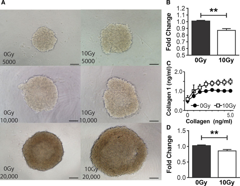
RT x alters the cellular functions of NHDF. A, NHDF spheroids imaged using bright-field microscopy (×10 objective) in the following variations: 0Gy 5,000 cells/spheroid, 10Gy 5,000 cells/spheroid, 0Gy 10,000 cells/spheroid, 10Gy 10,000 cells/spheroid, 0Gy 20,000 cells/spheroid, 10Gy 20,000 cells/spheroid validating the morphological increase in 10Gy NHDF size with the demonstrable increase in 10Gy spheroids size compared with the relevant 0Gy cells/spheroid control. B, Quantification of proliferation of NHDF demonstrates a significant reduction in proliferation after 10Gy irradiation compared with 0Gy controls at 48 h. C, Quantification of adherent 0Gy and 10Gy NHDF by solubilization of crystal violet and spectrophotometric analysis at 540 nm demonstrates a global increase in the number of adherent 10Gy NHDF at all collagen-I concentrations (0–5 ng/ml) compared with 0Gy controls. Fixed collagen-I gels were released circumferentially from well edges 48 h after seeding with 0Gy NHDF and 10Gy NHDF, allowed to float in media and contract over 24 h. D, Quantification of gel areas demonstrated a reduction in 10Gy NHDF collagen-I gel area, (reflecting increased contractile ability) compared with 0Gy NHDF gels. Scale bars (A) 100μm. (**P value < 0.01). Error bars represent SEM, n ≥ 3 for all experiments.