
Fig. 1.
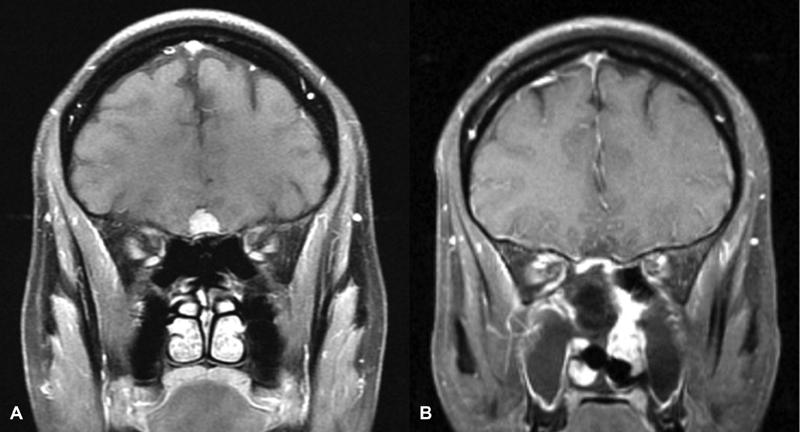
Patient 3 (A) preoperative T1 postcontrast MRI demonstrating a small unilateral OGM. (B) Postoperative T1 postcontrast MRI demonstrating interval resection of OGM and enhancing nasoseptal flap reconstruction. MRI, magnetic resonance imaging; OGM, olfactory groove meningioma.