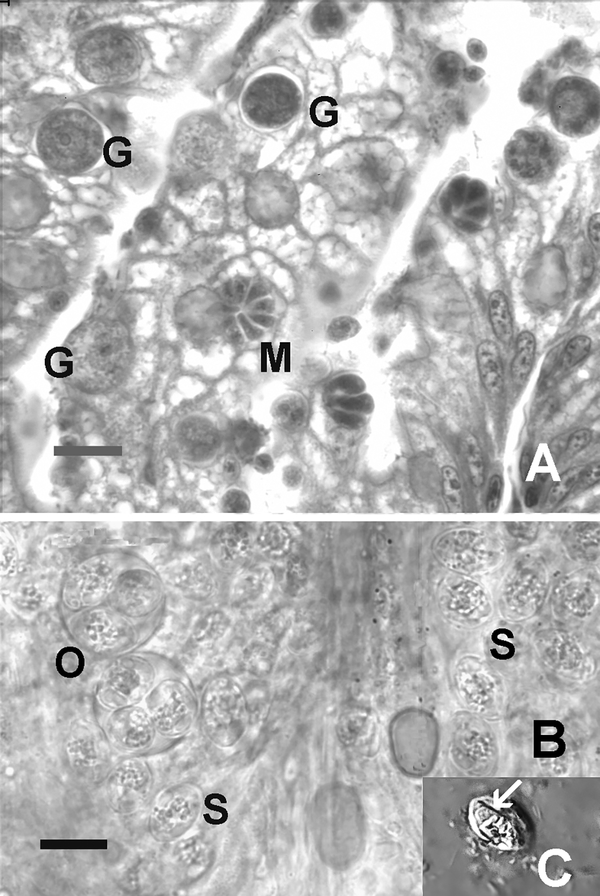
Figure 1.
Novel coccidian in zebrafish from a pet store. Bars = 10 μm A) Histological section of intestine showing various developmental stages in the epithelium. M = meront, G = gamont. Hematoxylin and eosin. B) Wet mount preparation (bright field) of coccidia in coelomic cavity. O = sporulated oocysts with four sporocysts. Numerous free sporocysts (S) are present. C) Sporocysts with suture (arrow), consistent with Goussia spp. Nomarksi phase interference.

