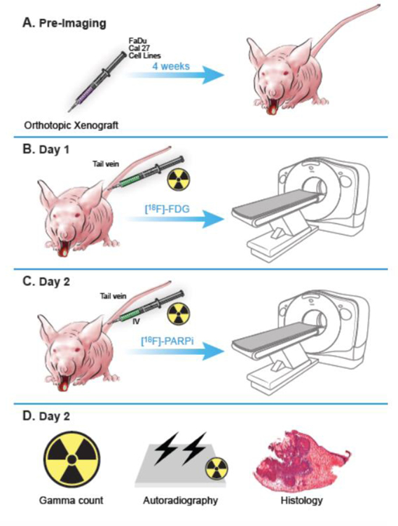
Fig 1.
Scheme of the study design and workflow. All animal experiments were performed in accordance with protocols approved by the Institutional Animal Care and Use Committee (IACUC) of MSK and followed the National Institute of Health guidelines for animal welfare. (A) Animals were inoculated on the anterior 1/3 and ventral portion of the right-hand side of the tongue with 500,000 cancer cells in 20 µL of PBS (n = 3 FaDu, n = 4 Cal 27), and tumors were allowed to proliferate for 4 weeks. (B) Mice were intravenously (tail vein) injected with an average of 7.7 ± 2.2 MBq (208.1 ± 59.4 µCi) of [18F]FDG on Day 1 after tumor establishment, and imaged 90 minutes after injection, under isoflurane anesthesia for 15 minutes. (C) The same animals were injected with an average of 10.4 ± 3 MBq (282.2 ± 80.6 µCi) of [18F]PARPi on Day 2, and imaged 90 minutes after injection. All animals were imaged on an INVEON small-animal micro-PET/CT scanner under isoflurane-induced anesthesia. (D) After [18F]PARPi imaging, animals were euthanized, and their tongues were harvested for radiation gamma-counting, autoradiography, and H&E staining.

