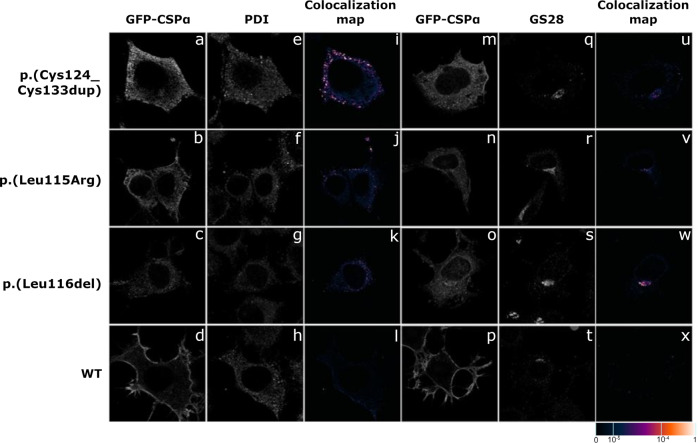
Fig. 2. Immunofluorescence analysis of transiently expressed GFP-tagged CSPα wt and variant proteins in CAD5 cells.
All three variant proteins (a–c, m–o) are present in a finely or coarsely granular structures. Co-staining with (e–g) protein disulfide isomerase (PDI), a marker of endoplasmic reticulum (ER), and (q–s) Golgi SNAP receptor complex member 1 (GS28) demonstrates abnormal presence of mutated proteins in ER (i–k) and Golgi (u–w). Wild-type protein (d, h, l, p, t, x) is present exclusively on plasma membrane. The degree of colocalization of GFP_CSPα with selected markers is demonstrated by the fluorescent signal overlap coefficient values ranging from 0 to 1. The resulting overlap coefficient values are presented as the pseudo color whose scale is shown in corresponding lookup table.

