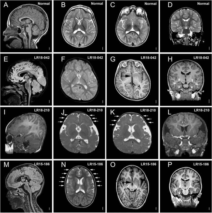
Fig. 1. Brain MRIs of individuals 1, 19 and 23 compared with MRIs of noncarrier TBR1 variants.
Brain MRIs of individuals with TBR1 variants affecting function in comparison with MRIs of noncarrier individuals (a–d) showing: g, o anomalies of the anterior commissure (AC) (black arrowheads); h, p hippocampal anomalies (white arrowheads); j, k, n cortical gyral anomalies (white arrows and black stars). LR18-042 = individual 1; LR15-186 = individual 23; LR18–210 = individual 19.

