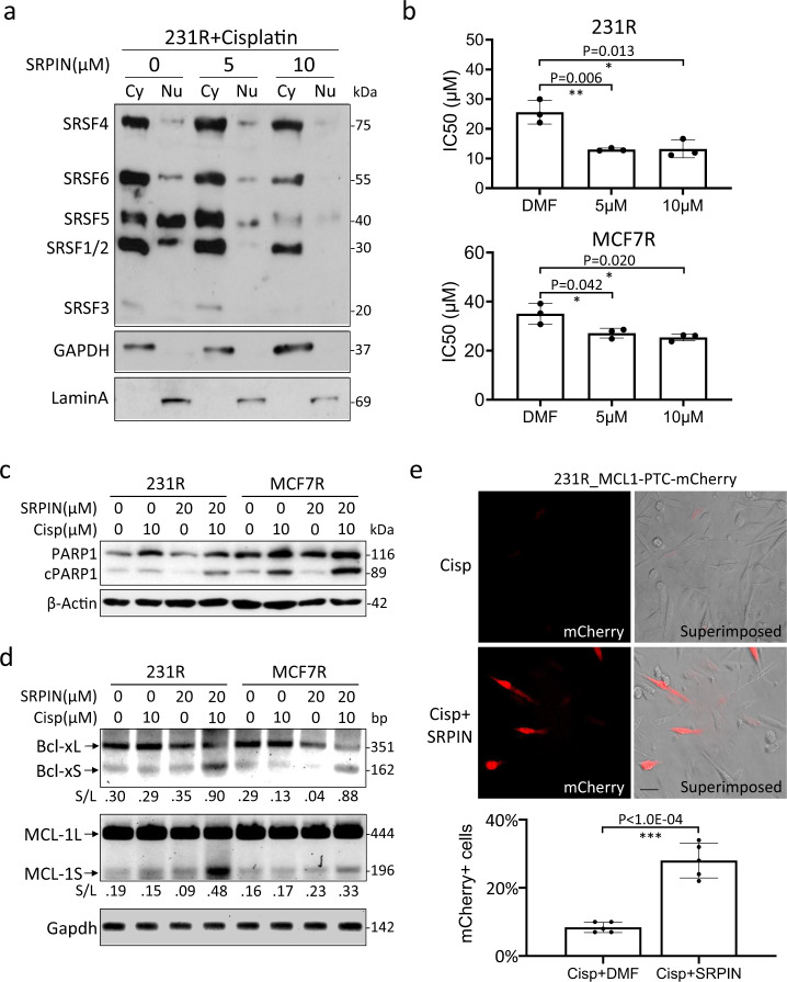
Fig. 8. Inhibition of the SRPK1 kinase activity sensitized cells to cisplatin.
a 231R cells were co-treated with 10 µM cisplatin and indicated concentrations of SRPIN340 (SRPIN). The cells were then fractionated into the cytoplasmic (Cy) and nuclear (Nu) portions for immunoblotting with mAb104. b 231R and MCF7R cells were treated with indicated concentrations of SRPIN340. The IC50 of cisplatin was then determined by MTS viability assays. Bars: mean ± SD; n = 3; *p < 0.05, **p < 0.01 as determined by Student’s t-test. c, d 231R and MCF7R were co-treated with cisplatin and SRPIN340 as indicated. The cleavage of PARP1 was assessed by immunoblotting (c), and the alternative splicing of BCL2L1 and MCL-1 by RT-PCR (d). The decimals below the gel strips in (d) denote the relative abundance of short (S) versus long (L) variants. e 231R cells were transfected with the splicing-sensitive reporter, MCL1-PTC-mCherry, and treated with cisplatin alone or together with SRPIN340. The mCherry signals were recorded by the fluorescence microscopy and superimposed onto the phase-contrast images. Scale bar: 20 µm. Bars: mean ± SD; n = 5; ***p < 0.01 by Student’s t-test. The Western blots in (a, c, d) are representative of three experiments with similar results.

