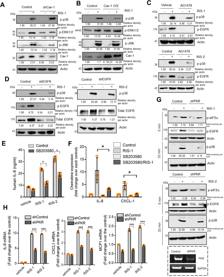
Fig. 5. Effects of Cav-1 on downstream signaling mediators of EGFR in ribosome-inactivated intestinal cells.
a HCT-8 cells transfected with control or shCav-1 vector were treated with vehicle or 50 ng/mL RIS-1 for 30 min. Cell lysates were subjected to western blot analysis. b HCT-8 cells transfected with control or Cav-1 overexpression vector were treated with vehicle or 50 ng/mL RIS-1 for 30 min. Cell lysates were subjected to western blot analysis. c HCT-8 cells were pre-exposed to control or 10 μM AG1478 for 2 h and treated with vehicle, 500 ng/mL RIS-2, or 50 ng/mL RIS-1 for 30 min. Cell lysates were subjected to western blot analysis. (d) HCT-8 cells transfected with control or shEGFR vector were treated with vehicle, 500 ng/mL RIS-2, or 50 ng/mL RIS-1 for 30 min. Cell lysates were subjected to western blot analysis. (e) HCT-8 cells were pre-exposed to vehicle or 10 μM SB203580 for 2 h and treated with vehicle, 50 ng/mL RIS-1, or 500 ng/mL RIS-2 for 12 h. The IL-8 concentration secreted into culture media was measured by ELISA. f HCT-8 cells were pre-exposed to vehicle or 10 μM SB203580 for 2 h and treated with vehicle, 50 ng/mL RIS-1, or 500 ng/mL RIS-2 for 1 h. IL-8 and CXCL-1 mRNA was measured using real-time RT-PCR. g HCT-8 cells transfected with control or shPKR vector were treated with vehicle, 500 ng/mL RIS-2, or 50 ng/mL RIS-1 for 5 or 30 min. Cell lysates were subjected to western blot analysis. h HCT-8 cells transfected with control or shPKR vector were treated with vehicle, 500 ng/mL RIS-2, or 50 ng/mL RIS-1 for 1 h. IL-8, CXCL-1, and MCP1 mRNA was measured using real-time RT-PCR. e–h Asterisks (*) indicate significant differences between groups (*p < 0.05, **p < 0.01, and ***p < 0.001 using two-tailed unpaired Student’s t test).

