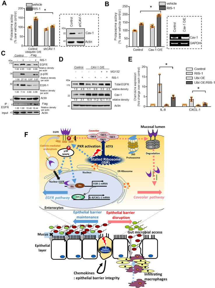
Fig. 7. Regulation of EGFR stability by Cav-1 in ribosome-inactivated intestinal cells.
a Control- or shCav-1-transfected HCT-8 cells were treated with vehicle or 50 ng/mL RIS-1 for 20 min and subjected to proteasome activity assay. b Control vector- or Cav-1-overexpressing HCT-8 cells were treated with vehicle or 50 ng/mL RIS-1 for 20 min; then, cells were subjected to proteasome activity assay. c Control- or Flag-tagged ubiquitin-overexpressing HCT-8 cells were treated with vehicle or 50 ng/mL RIS-1 for 20 min (total EGFR and Flag), 30 min (p-p38), and 1 h (Egr-1). Cell lysates were subjected to western blot analysis. Total lysates were immunoprecipitated with anti-EGFR antibody and subjected to western blot analysis. (d) Control- or Cav-1-overexpressing HCT-8 cells were pre-exposed to vehicle or 20 μM MG132 for 6 h and treated with vehicle or 50 ng/mL RIS-1 for 20 min. Cell lysates were subjected to western blot analysis. e Control- or Flag-tagged ubiquitin-overexpressing HCT-8 cells were treated with vehicle or 50 ng/mL RIS-1 for 1 h. IL-8 and CXCL-1 mRNA was measured using real-time RT-PCR. a, b, e Asterisks (*) indicate significant differences between groups (*p < 0.05, **p < 0.01, and ***p < 0.001 using two-tailed unpaired Student’s t test). Results and in vitro evaluations are representative of three independent experiments. f A putative scheme for Cav-1-mediated barrier disruption in the ribosome-inactivated gut. In response to ribosomal inactivation, ATF3-mediated Cav-1 production in caveolae counteracts the PKR- and EGFR-activated signaling pathways in intestinal epithelial cells. Activated caveolar clustering downregulates clathrin-dependent EGFR translocation and barrier-protective chemokine production. Disruption of PKR-EGFR-chemokine-mediated maintenance of barrier integrity allows microbial access into inner tissues, leading to further pro-inflammatory stimulation, including macrophage infiltration.

