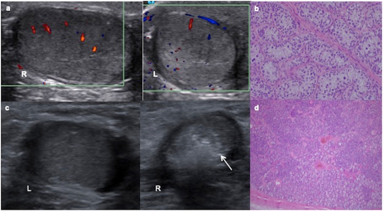
Fig. 9.
Two cases of intratubular germ cell neoplasia. a A 12-year-old boy with left cryptorchidism (inguinal testis). Colour Doppler ultrasound shows asymmetric size with normal right (R) testis and left (L) testis smaller, heterogenous and predominantly hyperechoic. b The surgical inguinal approach checks a hard cryptorchidic testis. Orchiectomy is practised and microscopic view of the pathologic specimen shows a large atypical cells with clear cytoplasm angulated nuclei with coarse chromatin, prominent nucleoli and cell borders resembling “fried egg” seminoma cells. c An 11-year-old boy with right cryptorchidism (inguinal testis). Ultrasound shows a normal left testis (L) and a small and heterogenous hyperechoic right testis (R) with small calcifications (arrow). d It is extirped giving rise to an intratubular germ cell neoplasia or carcinoma in situ of the testis: Spermatogenesis is absent and dystrophic calcifications are seen inside seminiferous tubules

