FIGURE 6.
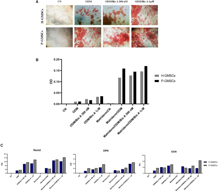
Osteoblastic differentiation assay. (A) Representative images of control H-GMSCs and P-GMSCs (P3) grown in osteogenic differentiation medium (ODM), with or without Biochanin A 300 nM and 1 μM, and stained with Red S Alizarin (4×); (B) histogram representing the quantitative analysis of Red S Alizarin by spectrophotometry (550 nm OD), of H-GMSCs and P-GMSCs (P3) grown in ODM, in presence or non-presence of the Matriderm® collagen scaffold, with or without Biochanin A 300 nM and 1 μM; (C) histogram showing the relative mRNA expression of the osteoblastic markers Runx2, OPN, and OCN in H-GMSCs and P-GMSCs (P3) grown in ODM, in presence or non-presence of the Matriderm® collagen scaffold, with or without Biochanin A 300 nM and 1 μM. Actin-β was used as the housekeeping gene; FC = fold change.

