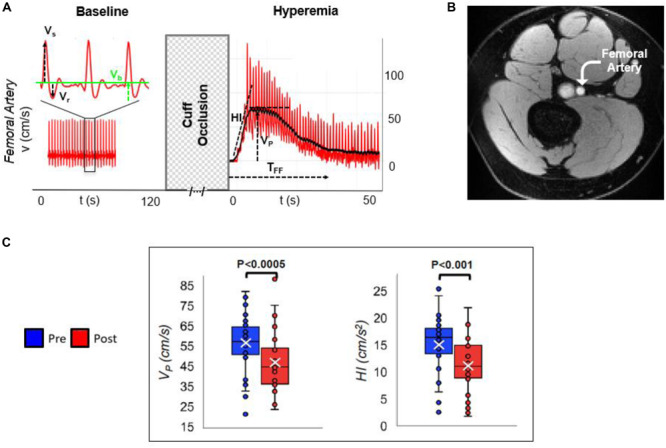
FIGURE 3.

Arterial velocimetry at baseline and hyperemia. (A) Blood flow velocity in the superficial femoral artery, during baseline and post-cuff occlusion (reactive hyperemia). Systolic, retrograde, and baseline velocities are indicated (Vs, Vr, and Vb, respectively). Post-ischemia, hyperemia is evaluated in terms of hyperemic index (HI) as the slope of the initial part of the velocity-time curve, peak average velocity (VP), and duration of hyperemia, referred to as time of forward flow (TFF). (B) Axial MR image of the thigh perpendicular to the femoral artery. (C) Pre- vs. post-electronic cigarette (e-cig) vaping differences for two parameters, in non-smokers, after a single episode of non-nicotinized e-cig vaping. The same MRI protocol was executed before and after e-cig use (modified from Caporale et al., 2019; Figures 2, 5, with permission from RSNA publisher).
