FIGURE 3.
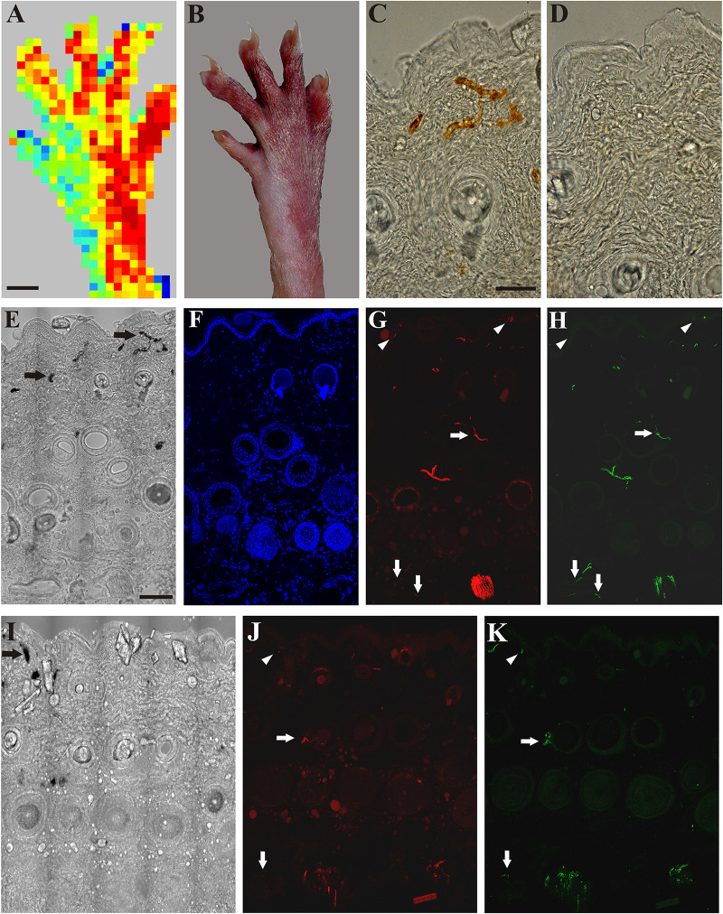
A: Application of mustard oil increased skin blood flow in the innervated (sciatic) but not in the denervated (saphenous) skin area of the right hind paw as assessed with scanning laser Doppler imaging. B: Brownish coloration of the skin due to silver accumulation in the innervated (sciatic) but not in denervated (saphenous) skin area of the dorsal hind paw skin elicited by mustard oil. C, D: Bright field photomicrographs showing sections from the lateral intact (C) and medial denervated regions of the hind paw skin after transection of the saphenous nerve (D). Silver-labeled small venules indicate increased vascular permeability of small postcapillary venules elicited by mustard oil application in the intact dorsal hind paw skin (C). Silver-labeled blood vessels cannot be observed in the denervated skin 4 days after saphenous nerve transection (D). E–H: Bright field (E) and immunofluorescence (F–H) photomicrographs showing the lateral (sciatic) skin area of the dorsal hind paw skin (E–H) after transection of the saphenous nerve. I–K: Bright field (I) and immunofluorescence (J, K) photomicrographs showing the medial (saphenous) skin area of the dorsal hind paw skin of a rat 15 days after transection and ligation of the saphenous nerve. Note the localization of ß-tubulin III (red) and CGRP-immunoreactive (green) nerve fibers in the epidermis (arrowheads), and around hair follicles and small arteries (arrows) in the innervated (lateral, sciatic) area of the dorsal hind paw skin identified by the presence of silver-labeled (permeable) venules (arrows in E). Fifteen days after transection and ligation of the saphenous nerve, some silver-labeled venules (arrow in I) and some ß-tubulin III and CGRP-immunoreactive epidermal (arrowheads in J, K) and dermal (arrows in J, K) nerve fibers can be observed in the medial (saphenous) skin area of the dorsal hind paw skin indicative of (collateral) regeneration. Scale bars indicate 5 mm in A and B and 50 μm in C and E. Scale bars in C and E apply for C, D and E–K, respectively.

