Figure 3.

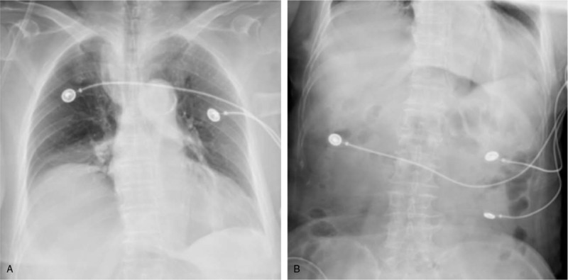
Chest (A) and semi-supine abdominal plain (B) radiographs showing enhanced lung markings, shadows in the left lower lung lobes, elevation of the right diaphragm, and mild pneumoperitoneum.

Chest (A) and semi-supine abdominal plain (B) radiographs showing enhanced lung markings, shadows in the left lower lung lobes, elevation of the right diaphragm, and mild pneumoperitoneum.