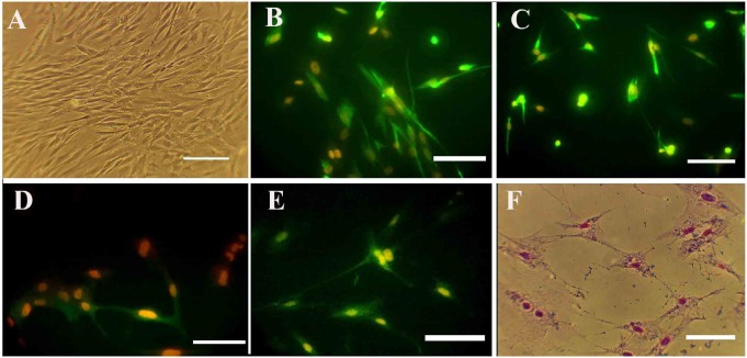
Figure 4.
Photomicrographs of hDPSCs for specific neural markers
A, B, C, D, E, and F. Represent the third passage, nestin, NF160, MAP2, ChAT, and cresyl violet staining, respectively. Nuclei were counterstained with propidium iodide (Scale bars: 50 μm).
hDPSCs: Human Dental Pulp Stem Cells; NF 160: Neurofilament 160; MAP2: Microtubule-Associated Protein 2

