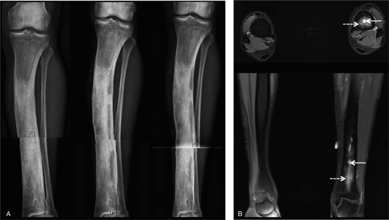
Figure 5.

Left distal tibia osteomyelitis. (A) Radiographic features (from left to right): initial X-ray, after surgical debridement and after 3 mo. (B) Axial and coronal magnetic resonance imaging, T1-weighted post contrast with fat suppression, note an intraosseous abscess (arrow) and marrow oedema (dashed arrow).
