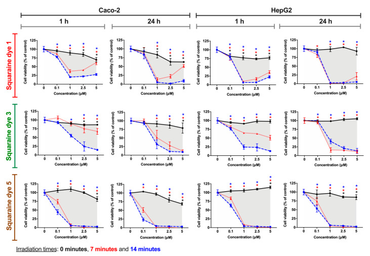
Figure 6.
Photodynamic effects of dicyanomethylene squaraine dyes 1, 3 and 5 on Caco-2 (human colorectal adenocarcinoma) and HepG2 (human hepatocellular carcinoma) cells. After cells’ incubation for 24 h with the respective dye (at 0.1, 1, 2.5 and 5 µM), cells were irradiated with suitable light-emitting diode systems for 0, 7, and 14 min (black, red and blue lines, respectively). After irradiation, the cells were maintained in contact with the irradiated dyes for 1 h or 24 h, and Alamar Blue reduction assay was performed for cell viability assessment. Results are expressed as % of control, non-treated cells, and presented as mean ± standard error of the mean (n = 3 independent experiments; each experiment in quadruplicates). Conditions with statistical significance compared to the negative control (p-value < 0.05) are indicated by an asterisk (*) of the color relative to the irradiation time. The shaded area delimits the difference in the percentage of cell viability between treatment with and without irradiation.

