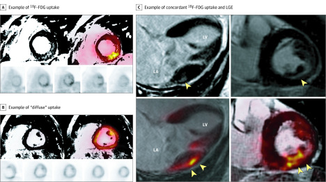
Figure 1. Hybrid Positron Emission Tomography/Magnetic Resonance Imaging (PET/MRI) of 3 Different Patients.
A, Example of fluorine 18–labeled fluorodeoxyglucose (18F-FDG) uptake that colocalizes with regions of late gadolinium enhancement (LGE) (PET+/MRI+) in a focal-on-diffuse pattern (maximum standardized uptake value [SUVmax], 3.5; maximum tissue-to-background ratio [TBRmax], 1.8; target-normal-myocardium ratio [TNMR], 1.8). These images were obtained from a man in his early 60s with asymptomatic degenerative mitral valve prolapse, significant mitral regurgitation (3+), and complex ventricular ectopy (nonsustained ventricular tachycardia and pleomorphic premature ventricular contractions) but without any objective surgical indications. The patient is being followed up with active surveillance. B, Example of diffuse uptake (SUVmax, 9.6; TBRmax, 4.2; TNMR, 1.3), which was interpreted as physiological or nonspecific uptake. C, Example of concordant 18F-FDG uptake and LGE in an asymptomatic patient with chronic severe degenerative mitral regurgitation and absent left ventricular remodeling (left ventricular ejection fraction, 60% and end-systolic dimension, 38 mm). The top 2 images are LGE imaging sequences and the bottom 2 images are 18F-FDG imaging. The yellow arrowheads indicate areas of either LGE or FDG uptake, respectively. LA indicates left atrial; LV, left ventricular.

