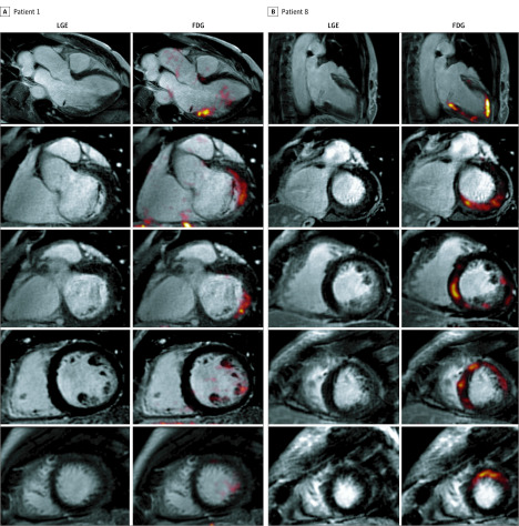
Figure 2. Representative Images of 2 Different Patterns of Fluorodeoxyglucose (FDG) Uptake on Hybrid Positron Emission Tomography/Magnetic Resonance Imaging (PET/MRI) From 2 Different Patients.
A, Focal FDG uptake and late gadolinium enhancement (LGE) in the inferolateral and basal inferior segments of the left ventricle in a man in his mid 50s with severe mitral regurgitation and posterior-leaflet prolapse who was asymptomatic with normal ventricular indices (left ventricular ejection fraction, 65%; left ventricular end-systolic dimension, 34 mm) and no objective indications for surgery. He developed episodes of nonsustained ventricular tachycardia (>15 beats) at peak exercise. B, Multifocal pattern of FDG uptake and LGE, in this case involving the septum, inferior, and lateral walls, in a woman in her mid 60s with symptomatic severe mitral regurgitation and bileaflet prolapse. She had infrequent premature ventricular contractions (burden = 0.5%), but they were pleomorphic (at least 3 different morphologies).

