
Figure 7.
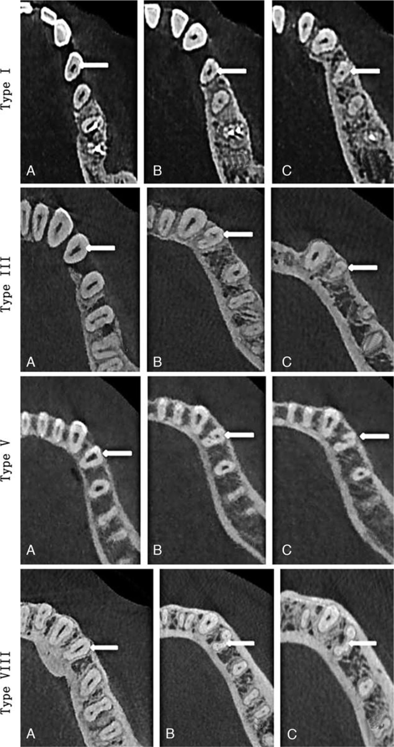
CBCT images (axial plane) showing various root forms of mandibular first premolars (arrows) at different levels of the root: (A) coronal; (B) middle third; and (C) apical third. CBCT = cone-beam computed tomography.
Official websites use .gov
A
.gov website belongs to an official
government organization in the United States.
Secure .gov websites use HTTPS
A lock (
) or https:// means you've safely
connected to the .gov website. Share sensitive
information only on official, secure websites.
CBCT images (axial plane) showing various root forms of mandibular first premolars (arrows) at different levels of the root: (A) coronal; (B) middle third; and (C) apical third. CBCT = cone-beam computed tomography.