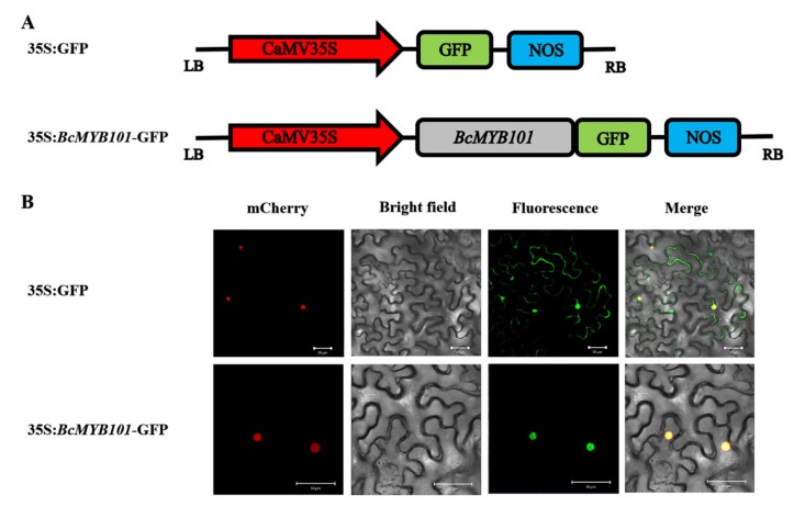
Figure 4.
Subcellular localization of BcMYB101. (A) The construct of 35S:GFP (green fluorescent protein) and 35S:BcMYB101-GFP fusion protein. (B) The panels from left to right correspond to the mCherry (nuclear marker), bright-field, fluorescence, and merged fluorescence images of 35S:GFP and 35S:BcMYB101-GFP fusion protein. Scale bars = 50 µm.

