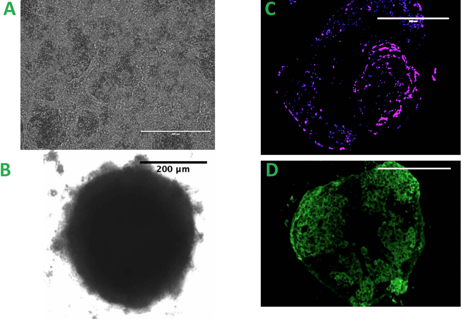
Figure 1.

A. iPSC differentiated Cardiomyocyte, scale bar = 400μm B. The spheroid created from the co-culture of three types of cells, scale bar = 200 μm C&D. Spheroids with CD31 positive staining C. The magenta: the presence of endothelial cell, scale bar = 200 μm D. The green: Troponin T staining of individual spheroid, scale bar = 200 μm
