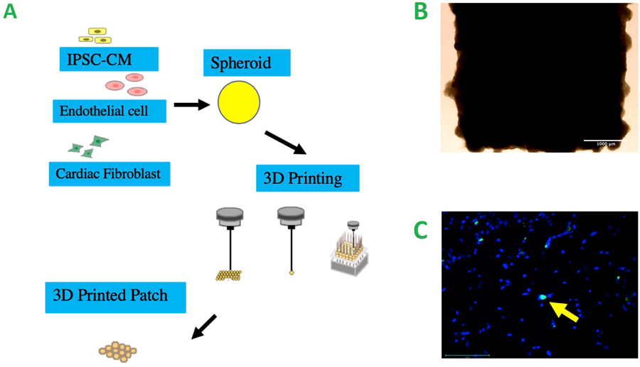
Figure 2.

A. Schematic of bioprinting, co-cultures of the cells for the creation of spheroids, 3D bioprinting of the cardiac patch from the spheroids B. Cardiac patch from the bioprinting prior to implantation, scale bar = 1000 μm C. TUNEL staining of the 3D printed cardiac patch 4 weeks after fabrication, Yellow arrow: apoptotic cells. scale bar = 100 μm
