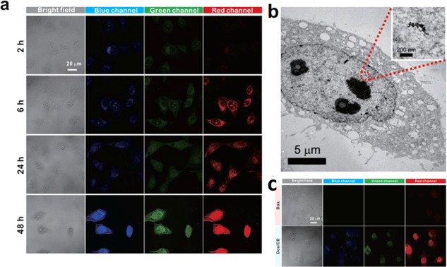
Figure 3.
Nucleus staining of HeLa cells with zwitterionic CDs. (a) Bright-field and confocal fluorescence microscopy images for different incubation times to show the cytoplasmic and nuclear transport of CDs. (b) Biotransmission electron microscopic image showing localization of CDs in the nucleus. (c) Bright-field and confocal fluorescence images of HeLa cells treated with DOX and DOX/CD. Reproduced with permission from ref (29). Copyright 2015 Springer Nature.

