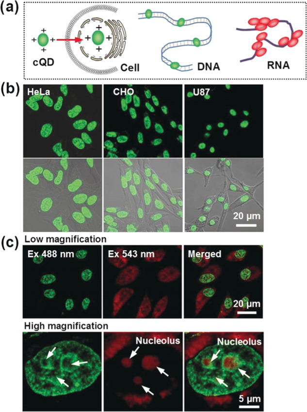
Figure 5.

(a) Schematic of CDs binding to DNA and RNA. (b) Fluorescence images of different cell lines treated with CDs for nucleus imaging. (c) Multicolor fluorescence imaging of HeLa cells with the CDs using multiple excitation and emission wavelengths. Reproduced with permission from ref (70). Copyright 2019 Wiley-VCH.
