
Fig. 7. Probing dock-lock mechanism in Aβ1–40 by solution NMR.
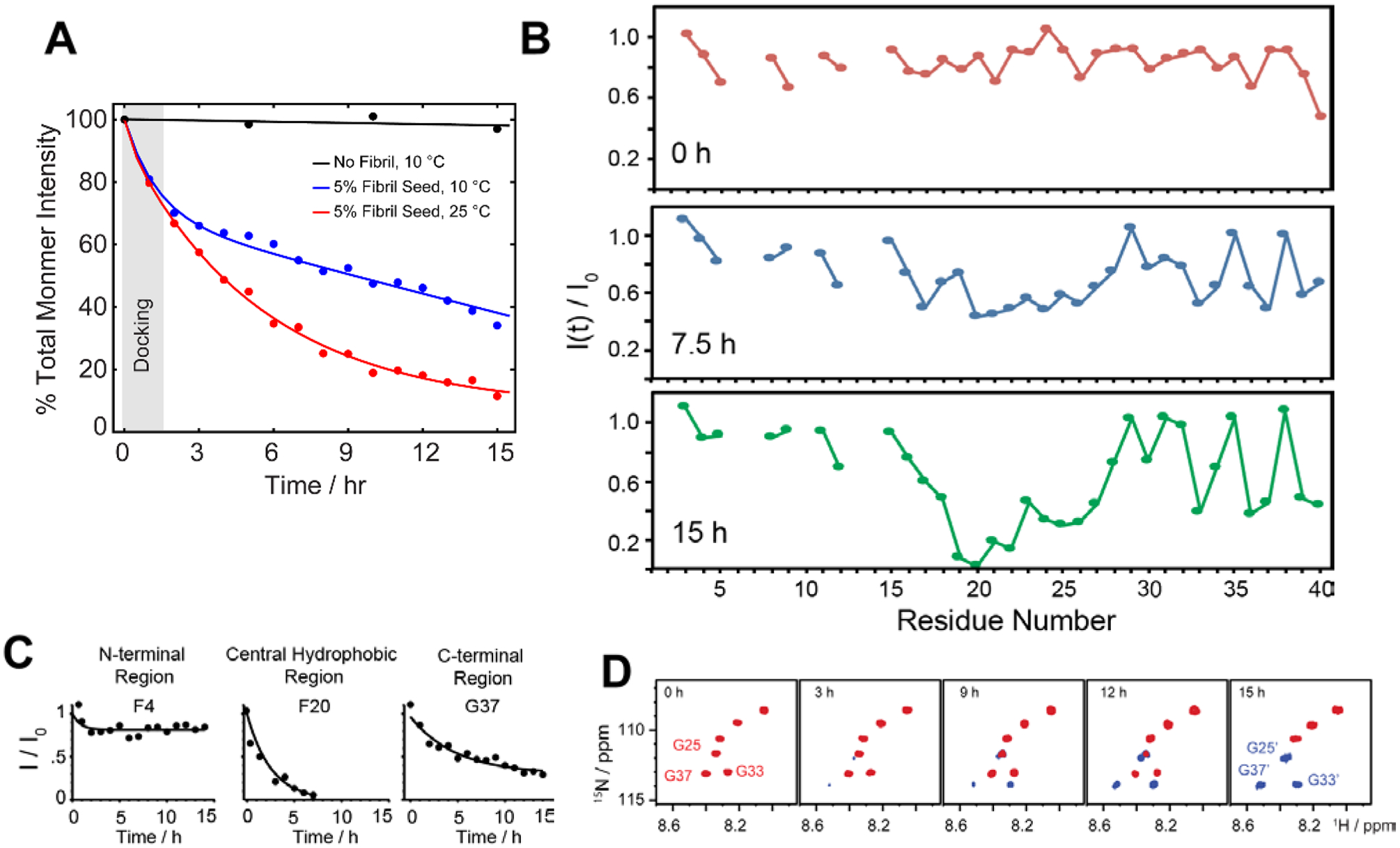
(A) Monitoring the depletion of total intensity obtained from 2D 1H-15N SOFAST-HMQC spectra during a self-seeding reaction at 10 °C. The observed distinguished kinetic phase (black vs blue curve) indicates the dominant docking phase (grey shade) within a time-scale of first couple of hours. (B–C) Time-interval measurement highlights a substantial drop in NMR signal intensities of the central hydrophobic residues (F20 as a representative residue) as compared to N- or C-terminal residues (F4 and G37 as representative residues) indicating a possible docking site in monomer onto fully matured fibers. (D) NMR self-seeding reaction identified appearance of new peaks in the SOFAST-HMQC spectrum indicating the origin of new oligomer species. Copyright © 2019 Royal Society of Chemistry. Reproduced by permission from the Royal Society of Chemistry https://doi.org/10.1039/C9CC01067J. Further details are available in the referenced work.116