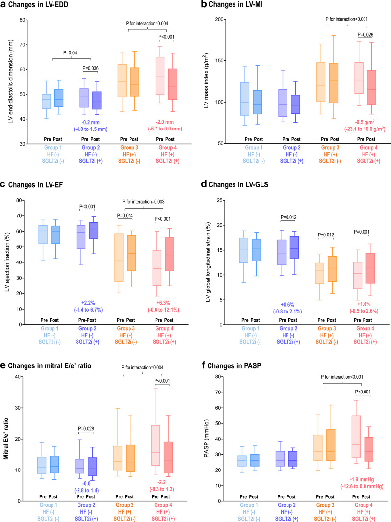
Fig. 2.
Changes in LV function and geometry by SGLT2i according to the presence of HF and the use of SGLT2i. Echocardiographic parameters at baseline and follow-up are presented according to the presence of HF and the use of SGLT2i: a LV-EDD, b LV-MI, c LV-EF, d LV-GLS, e mitral E/e′ ratio, and f PASP. Bars represent the median with interquartile range (Q1–Q3). Intra-group and inter-group comparisons were performed with paired t-test generalized linear model for repeated measure analysis, respectively. LV left ventricular, EF ejection fraction, GLS global longitudinal strain, EDD end-diastolic dimension, MI mass index, PASP pulmonary arterial systolic pressure; others as in Fig. 1

