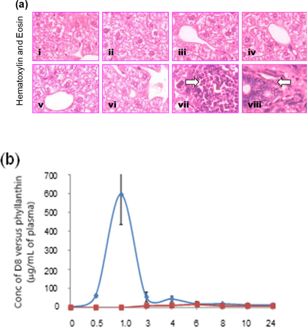
Figure 9.

(a) Histopathological examination of the mice liver. A representative picture from the different groups (1–4) in the acute toxicity studies, (i) water control; (ii) D8 4000 mg/kg b. wt; (iii) PEG-400 (300 μL/mouse); (iv) phyllanthin (1500 mg/kg b. wt.). Similarly, the groups (5–8) in the subacute toxicity study, (v) water control; (vi) D8 (1000 mg/kg b. wt/day); (vii) PEG-400 (200 μL/day/mouse); (viii) phyllanthin (500 mg/kg b. wt/day). Arrow indicates multifocal aggregation of hematopoietic cells and liver necrosis. (b) Bioavailability of D8 (500 mg/kg b. wt p.o) and phyllanthin (1500 mg/kg b. wt p.o) in BALB/c mice. Values used to plot AUC are mean ± SD from the three independent experiments.
