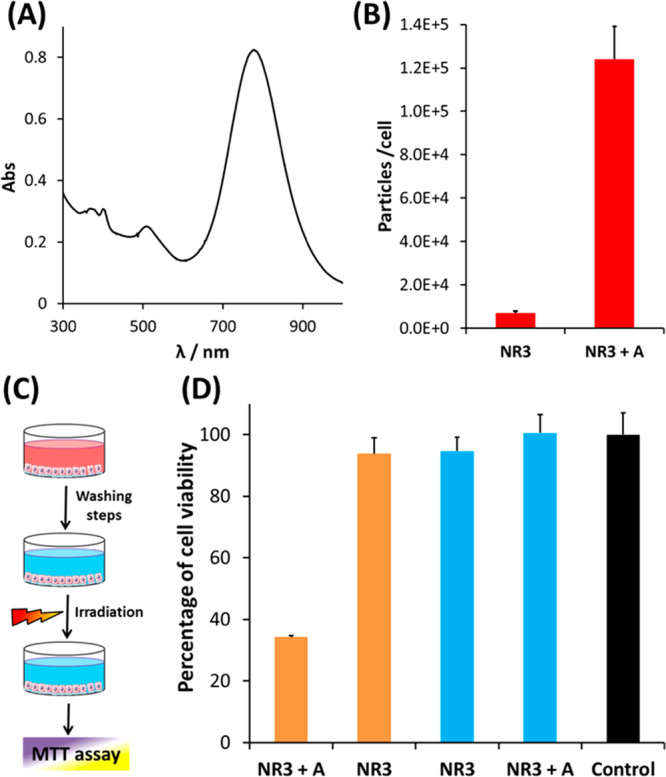
Figure 5.

(A) UV–Vis–NIR spectrum of NR3. (B) Cell uptake of NR3 (9 × 10¹⁰ NR/mL) by HeLa cells, in the absence and in the presence of cage A (5 μM), as determined by ICP-MS. The incubation was performed in DMEM medium with 10% FBS for 24 h. (C) Schematic representation of the protocol followed in the NIR-laser hyperthermia experiments. HeLa cells were first incubated with NR3 (9 × 10¹⁰ NR/mL) for 24 h in DMEM medium with 10% FBS, with or without 5 μM cage A. Subsequently, noninternalized nanorods were removed by washing with PBS, and cells were irradiated with an 808 nm diode laser (Lumics, LU808T040) for 20 min at a power density of 3.2 W/cm2. Cell viability was calculated using the MTT assay (average of triplicate wells ± standard deviation). (D) Results of the irradiation experiment. The control bar (black) represents cell viability of the cells in the absence of cage A and NR3. Blue bars represent incubation without irradiation, proving that uptaken NR3 in the presence of cage A did not induce cytotoxicity.
