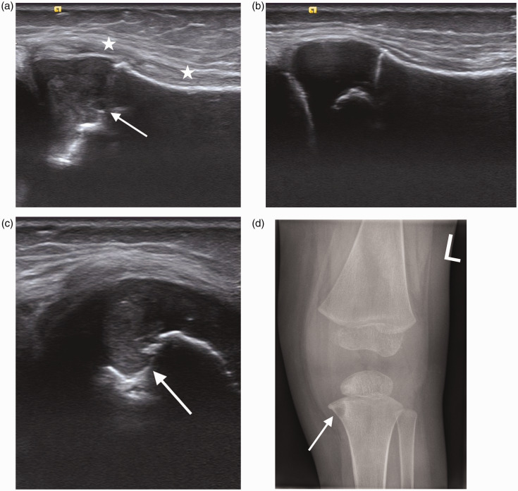
Figure 5.
Two year old with tibial osteomyelitis. (a) Longitudinal scan of the left proximal tibia shows increased echogenicity in the metaphysis and epiphysis (arrow). Marked superficial soft tissue thickening (*) is best appreciated when compared to the normal proximal right tibia in (b). (c) Transverse scan of the proximal tibia demonstrates an echogenic focus and bone destruction in the metaphyseal region (arrow). (d) AP radiograph of the left knee shows the same area of bone destruction (arrow).

