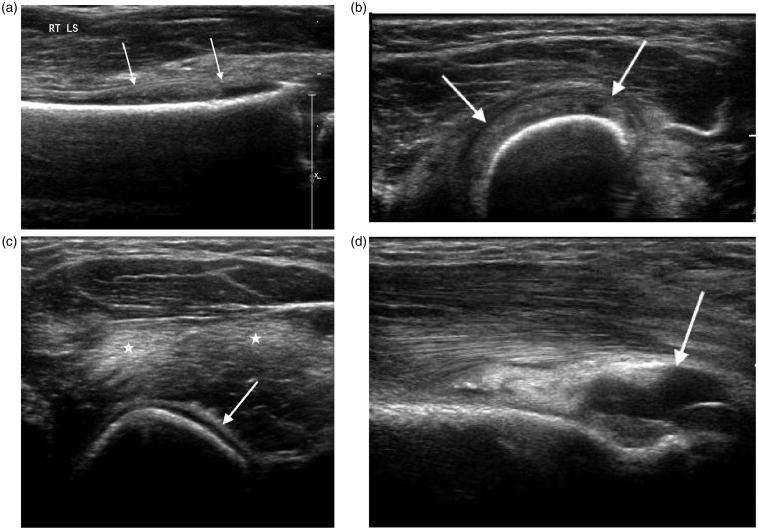
Figure 8.
Two year old with humeral osteomyelitis. (a) Longitudinal and (b) transverse scans of the distal humerus demonstrate an echogenic subperiosteal collection (arrows). (c) Transverse scan of the upper arm demonstrates periosteal reaction (arrow) and associated muscle oedema (*). (d) Longitudinal scan of the posterior elbow shows an associated joint effusion (arrow).

