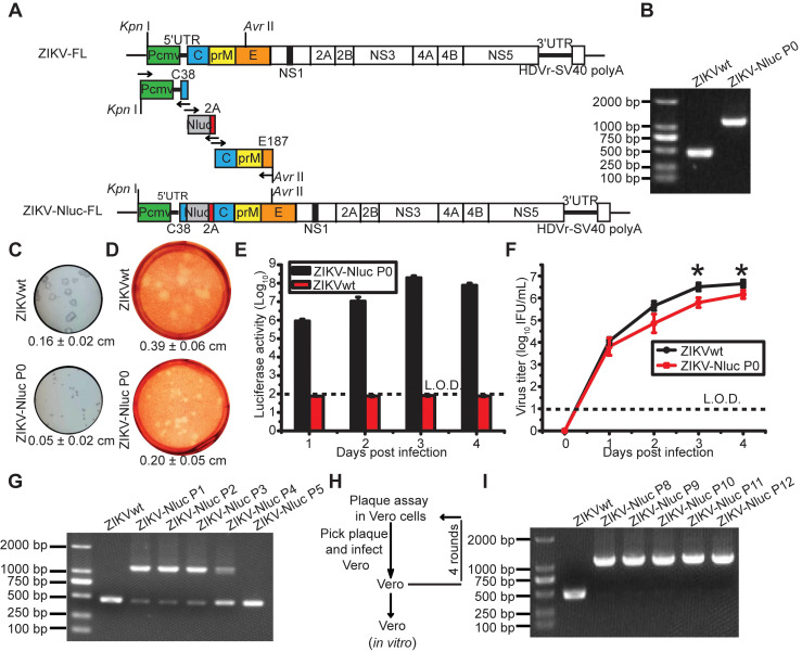
Figure 1.
Generation and characterization of ZIKV-Nluc. (A) Strategy for constructing the full-length cDNA clone of ZIKV-Nluc. The monomeric Nluc gene flanked by the N-terminal 38 amino acids of C protein (C38) and a FMDV2A (F2A) sequence was inserted between 5'UTR and the C gene. (B) The stability of the P0 ZIKV-Nluc virus. Viral RNAs were extracted from the supernatants, and RT-PCR was performed with a pair of primers surrounding the Nluc-2A fragment. (C) The plaque morphology of the P0 ZIKV-Nluc virus in Vero cells, visualized by immunostaining following incubation for 4 days. (D) The plaque morphology of the P0 ZIKV-Nluc virus in Vero cells, visualized using 0.33% neutral red following incubation for 5 days. (C, D) The average sizes of viral plaques (mean ± standard deviation) were quantified by counting all of the intact plaques. (E) Nluc activities of the infected Vero lysates by P0 ZIKV-Nluc virus at different times post infection at low multiplicity of infection (MOI) of 0.01. (F) Growth curves of P0 ZIKV-Nluc virus determined by an immunostaining plaque assay at an MOI of 0.01. (G) ZIKV-Nluc stability during virus passaging. Total RNA was extracted from the cells infected by each passaged virus, and RT-PCR was performed with a pair of primers surrounding the Nluc-2A fragment. (H) Schematic of plaque purification. (I) ZIKV-Nluc stability following plaque purification. Data represent the mean ± SD analysed by Student's t-test (two tailed) (*, P < 0.05; **, P < 0.01; ***, P < 0.001).

