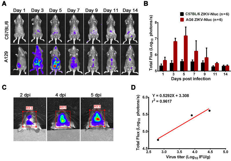
Figure 4.
In vivo luminescence of ZIKV-Nluc-infected mice. (A, B) Groups of A129 and C57BL/6 mice (3-4 weeks old; n = 6) were infected intraperitoneally with 1.2 × 10⁵ IFU of WT or ZIKV-Nluc. (A) Bioluminescence imaging of ZIKV-Nluc-infected mice was performed at the indicated times. Representative ventral views of the results were shown. (B) The average radiance of ZIKV-Nluc-infected mice was determined from region of interest (ROI) analysis of the ventral side. (C, D) Groups of AG6 mice (3-4 weeks old; n = 3) were infected with 6×10³ IFU of ZIKV-Nluc via the i.c. route. (C) Bioluminescence imaging of ROI from ZIKV-Nluc-infected mice was performed at the indicated times. (D) Linear correlation between the viral titres and Nluc signal values of the ZIKV-Nluc virus in vivo.

