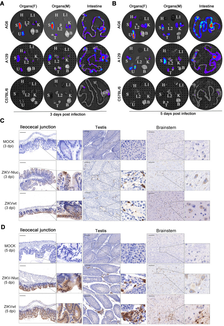
Figure 8.
Tissue localization of ZIKV-Nluc. Groups of AG6, A129 and C57BL/6 mice (3-4 weeks old; n = 6) were infected with 6 ×10⁴ IFU of WT or ZIKV-Nluc via footpad. Immediately after bioluminescence imaging, ZIKV-Nluc-infected mice were sacrificed, and isolated organs including heart (H), liver (L1), spleen (S), lung (L2), kidney (K), uterus/ovary (U), testis (T), and brain (B) were subjected to in vitro bioluminescence imaging at 3 dpi (A) and 5 dpi (B). The expressions of E protein in ileocecal junction, testis and brainstem sections from infected AG6 mice were stained by immunohistochemistry at 3 dpi (C) and 5 dpi (D). Scale bars are 100 μm for lower magnification images (× 20) and boxes in lower magnification images indicated where the higher magnification images (× 80) were taken.

