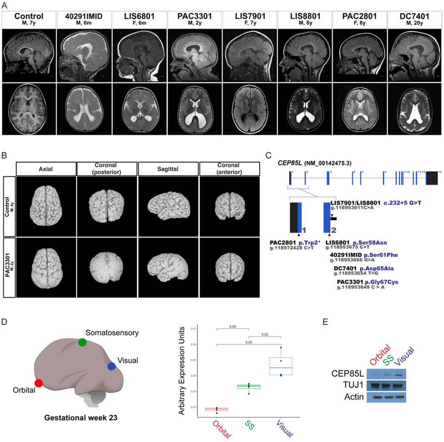
Figure 1: Variants in CEP85L cause posterior-specific pachygyria.

A. Sagittal and axial plane MRI images of a control and affected individuals with posterior reduced gyral folding. B. Three-dimensional MRI presentation of a control and PAC3301 patient with a de novo CEP85L variant. C. Schematic representation of exons of CEP85L shown as blue bars. The variants in CEP85L are found in exons 1 and 2. D. Brain region-specific qPCR of gestational week 23 cortex (GW), demonstrating the increasing rostral-to-caudal expression pattern of CEP85L normalized to β-actin. Orbital (red), somatosensory (green), and visual (blue) cortex. For quantifications, one brain region was analyzed in triplicate or quadruplicate (n=1). p < 0.03 (Student T-test). E. Whole cell lysate from the posterior frontal, parietal and occipital lobes of a GW 23 fetus blotted for CEP85L and the lissencephaly-associated protein, LIS1. Actin and TUJ1 served as a loading control and neuron-specific sampling control, respectively.
