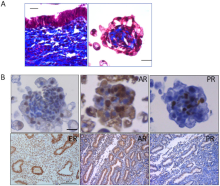
Figure 3: Endometrial organoids exhibit characteristics of native tissue after 14 days of follicular phase hormone treatment.
(A) Trichrome staining was done to visualize collagen (blue) and cells (red). Trichrome stain was performed on endometrial tissue (left panel) and endometrial organoids (Scale bars = 20 μm, right panel). (B) Immunohistochemical staining for ER, AR and PR was done in endometrial tissue (Scale bar = 100 μm bottom panels) and endometrial organoids (Scale bar = 20 μm, top panels). Positive stain is shown in brown and hematoxylin stain solution was added as the counter stain shown in blue. Adapted from Teerawat et al., JCEM, (2019)11.

