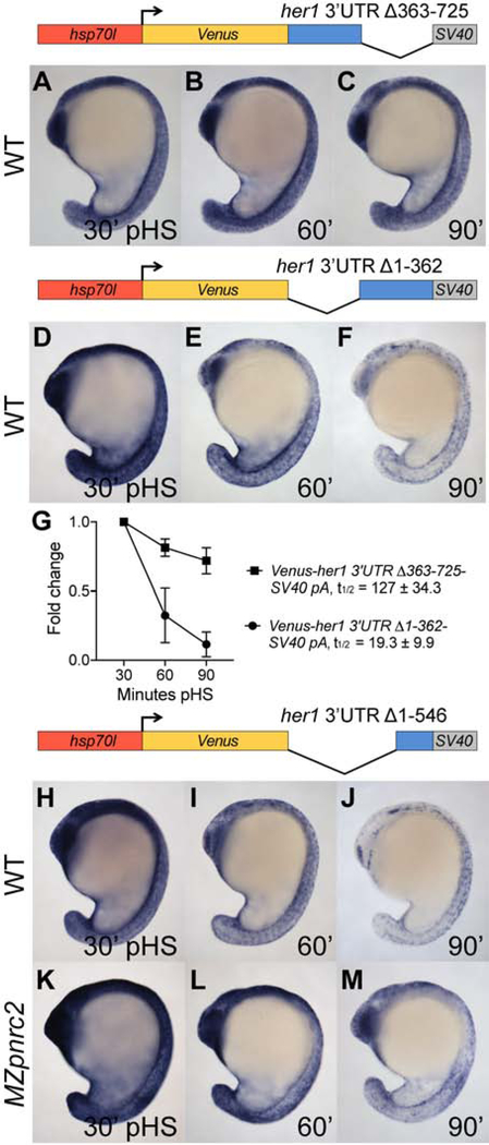
Figure 3. The terminal 179 nucleotides of the her1 3’UTR is sufficient for Pnrc2-mediated decay of reporter transcripts.
(A–F) Transgenic embryos carrying the hsp70l:Venus-her1 3’UTRΔ363–725-SV40 pA reporter (line oz54) or hsp70l:Venus-her1 3’UTRΔ1–362-SV40 pA reporter (line oz47) were raised to mid-segmentation stage and heat shocked for 15 minutes, then collected at the indicated minutes pHS and processed by Venus in situ hybridization (n ≥ 6 per time point). Venus transcript is not detected in the absence of heat shock (n = 10 per reporter line) (data not shown). (G) qPCR analysis comparing Venus transcript fold change from 30 minutes pHS to 60 and 90 minutes pHS for the Tg(hsp70l:Venus-her1 3’UTRΔ363–725-SV40 pA)oz54 or Tg(hsp70l:Venus-her1 3’UTRΔ1–362-SV40 pA)oz47 reporter lines (n = 10 embryos per time point across three biological replicates). (H–M) Mid-segmentation stage wild-type (WT) and MZpnrc2 mutant embryos carrying the hsp70l:Venus-her1 3’UTRΔ1–546-SV40 pA reporter (line oz50) were heat shocked and processed by Venus in situ hybridization (n ≥ 7 embryos per time point). Venus transcript is not detected in the absence of heat shock (n = 10 wild-type embryos) (data not shown). Representative embryos were genotyped post-imaging to confirm genotype. For each reporter, three independent lines were analyzed in wild-type embryos by in situ hybridization and exhibited comparable Venus decay across all lines carrying the same reporter (data not shown); one representative line for each is shown (see Methods for details). pHS = post-heat shock; t1/2 = half-life; ± = standard deviation; pA = polyadenylation sequence.

