Figure 2.
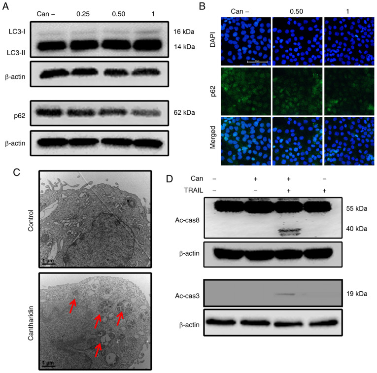
Can induces autophagy and enhances TRAIL-mediated apoptosis of cells. DU145 cells were treated with Can (0, 0.25, 0.50 and 1 μM) for 18 h. (A) The levels of LC3-II and p62 were assessed by western blot analysis. (B) DU145 cells were treated with Can (0, 0.50 and 1 μM) for 18 h. Cells were then immunostained with p62 (green) and evaluated for fluorescence (scale bar=50 μm; magnification, ×200). (C) The formation of autophagosomes in treated cells was examined by transmission electron microscopy. Red arrows indicate autophagosomes. Scale bar=1 μm. (D) DU145 cells were treated with Can (1 μM) for 18 h and then TRAIL for an additional 1 h. The levels of Ac-cas3 and Ac-cas8 were assessed by western blot analysis. β-actin was used as the control. TRAIL, tumor necrosis factor-related apoptosis-inducing ligand; can, cantharidin; Ac-cas, activated caspase; p62, sequestosome 1; LC3-I, cytoplasmic microtubule-associated proteins 1A/1B light chain 3B; LC3-II, lipid-modified microtubule-associated proteins 1A/1B light chain 3B.

