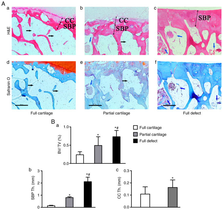
Figure 2.
Alternation of the microstructure in subchondral bone from human knee. (A) H&E staining of the (a) full, (b) partial cartilage and (c) full defect. (Ad-f) Safranin O/fast green staining in (d) full, (e) partial cartilage and (f) full defect groups. In the full cartilage group, the bone marrow cavity was mainly filled with adipose tissue (black arrow), but in the partial cartilage and especially in full defect groups, it was composed of bone marrow cells (blue arrow), in which new trabecular bone formed locally (red color in H&E and blue in safranin O staining). Quantitative results showing that subchondral bone possesses a (Ba) higher BV/TV fraction, (Bb) SBP Th. and CC Th. in cartilage wear groups, as compared with (Bc) the full cartilage group. *P<0.05 vs. the full cartilage and #P<0.05 vs. the partial cartilage groups, n=21. Scale bar, 500 µm. H&E, hematoxylin and eosin; BV/TV, Bone volume/total tissue volume; CC Th., Thickness of calcified cartilage; SBP Th., Thickness of subchondral bone plate.

