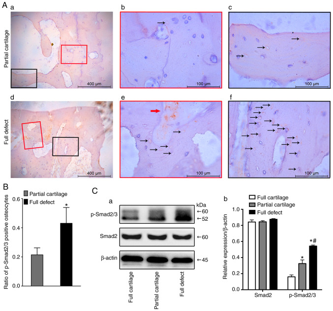
Figure 4.
Activation of osteocyte TGFβ signaling in human subchondral bone. The activation of osteocyte TGFβ signaling was detected by immunohistochem-istry staining with phosphorylated smad2/3 antibody in (Aa-c) partial cartilage defects and (Ad-f) full defect groups. (Ab) Magnification of the super-layer structure in subchondral bone of Aa. (Ac) Magnification of the deep-layer structure in subchondral bone of Aa. (Ae) Magnification of the super-layer structure in subchondral bone of Ad. (Af) Magnification of the deep-layer structure in subchondral bone of Ad. (B) Quantitative assay was performed by ratio of p-Smad2/3+ osteocytes. Positive cell stained brown in osteocytes (black arrow) and bone marrow (red arrow). (Ca) Western blotting was performed to test the activation of the TGFβ-Smad2/3 signaling pathway in subchondral bone from three different groups and (Cb) quantitative analysis for the relative expression of Smad2 and p-Smad2/3 was performed. *P<0.05 vs. the full cartilage and #P<0.05 vs. the partial cartilage groups n=21. Scale bar in Aa and d, 400 µm; in others, 100 µm. TGFβ, transcriptional growth factor β; p, phosphorylated.

