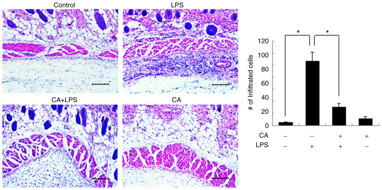
Figure 7.
CA inhibits inflammatory cell infiltration in vivo. Rats were intraperitoneally injected with CA. After 3 h, LPS was subcutaneously inoculated into the right flanks of the rats. After 24 h, subcutaneous tissues were excised and stained with H&E. Inflammatory cell infiltration was evaluated by light microscopy. Original magnification, ×100; scale bar, 10 µm. The graph shows the quantitative evaluation of the infiltrated cells. The data are expressed as the means ± SD of 3 individual experiments. *P<0.05. CA, cinnamaldehyde; LPS, lipopolysaccharide.

