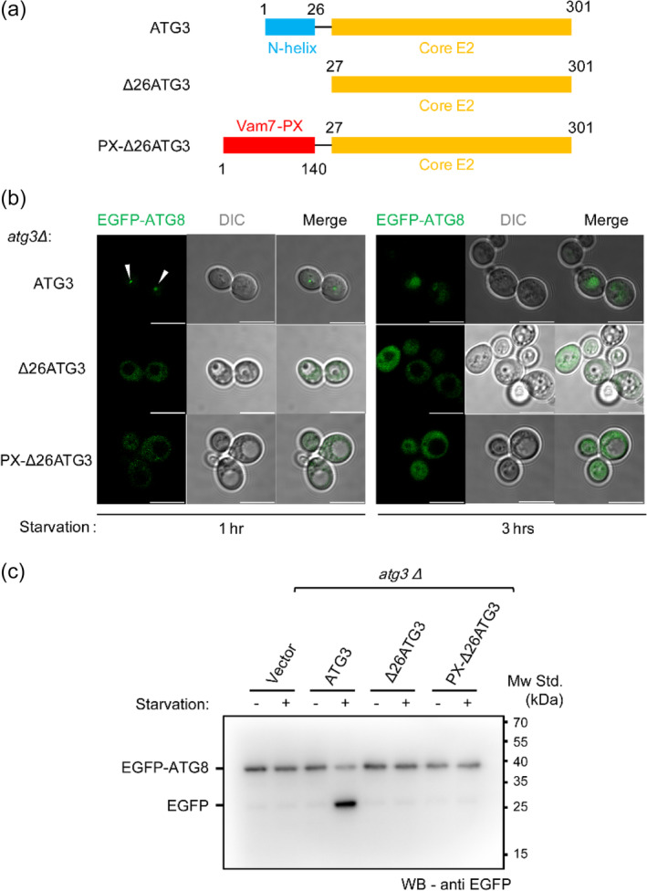
FIGURE 1.

The N‐terminal amphipathic helix of Atg3 is required for autophagy initiation. (a) Illustration of the domain structures of Atg3 and variants used in this study. The N‐terminal helix and core E2 domain of Atg3 are colored with blue and orange, respectively. The genetically engineered PX domain of Vam7 (residues 1–140) is colored with red. (b) Representative laser scanning confocal microscopy (LSCM) graphs of yeast atg3Δ cells expressing EGFP‐Atg8, Atg3, and its variants upon 1‐ and 3‐hr starvation. The white arrows indicate EGFP‐Atg8 puncta during autophagy initiation. Scale bars, 10 μm. (c) EGFP‐Atg8 processing assay as shown by anti‐EGFP immunoblotting from the lysates of yeast atg3Δ cells expressing EGFP‐Atg8, Atg3, and its variants. The cells were grown to mid‐log phase and either harvested (−) or nitrogen starved for 3 hr (+). The degraded EGFP after starvation represents normal autophagy flux
