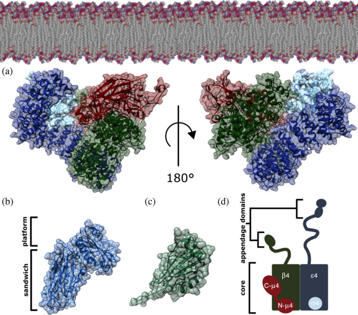
FIGURE 1.

AP‐4 homology model. (a) AP‐4 core in its open conformation is depicted with ε, β4, μ4, and σ4 subunits shown in blue, green, red, and cyan, respectively. The complex is shown in the orientation thought to interact with the membrane, as based on related AP structures. The AP‐4 core homology model was generated using existing experimental structures of both AP‐2 (PDB: 2XA7) and AP‐4 (PDB: 3L81). (b) AP‐4 ε appendage domain homology model showing the platform and sandwich subdomains generated from AP‐2 α appendage (PDB: 1B9K). (c) β4 appendage domain (PDB ID: 2MJ7). (d) Schematic of the AP‐4 heterotetramer. The ε and β4 subunits contain C‐terminal appendage domains (20–30 kDa each) attached to the core (200 kDa) via long unstructured linkers. AP‐4, adapter protein complex 4
