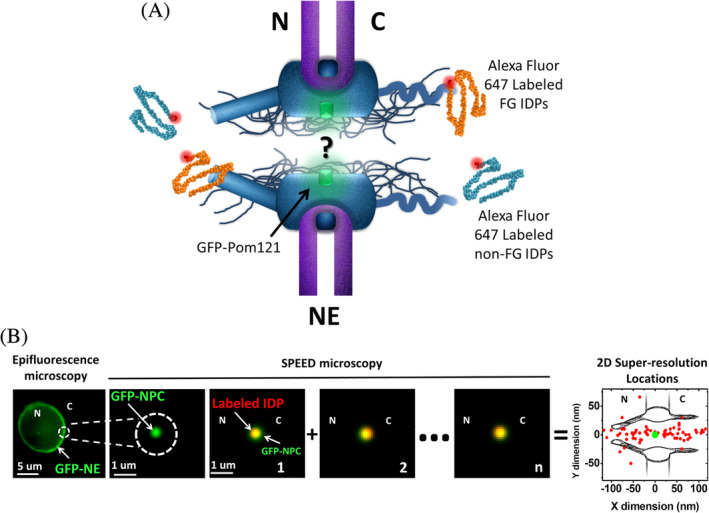
Figure 1.

Nuclear transport of IDPs through the NPC illuminated by SPEED microscopy. (a) The fundamental mechanism for nuclear transport of IDPs through the NPCs remains unknown. Native NPCs embedded into the NE were labeled through genetically tagging green fluorescent protein (GFP) to a scaffold Nup POM121, in which the fluorescence of GFP‐POM121was utilized to localize the NPC. Alexa Fluor 647 dye molecules were used to label FG‐ and non‐FG‐IDPs. N, nucleus; C, cytoplasm. (b) Wide‐field epifluorescence image shown a green fluorescent ring of NE in a permeabilized HeLa cell expressing GFP‐POM121. SPEED microscopy illuminated only a single NPC on the NE. Labeled IDPs were tracked as they diffused through the NPC and their dwelling positions (red dots) were localized and then superimposed to obtain a 2D super‐resolution distribution around the NPC centroid (green dot). N, nucleus; C, cytoplasm. IDP, intrinsically disordered protein; NE, nuclear envelope; NPC, nuclear pore complex; SPEED, single‐point edge‐excitation sub‐diffraction
