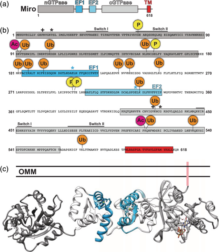
Figure 1.

Structural features of Miro. (a) Domain organization. (b) Annotated primary sequence of Miro. Black “+” indicates constitutively active GTP mutant whereas “*” indicates constitutively inactive GTP mutant. Blue “*” indicate mutations in EF‐hands that block calcium binding. (c) Model of Miro structure based on published coordinates of Miro's nGTPase (PDB: 6D71) and the C‐terminus of Miro (PDB: 5KSZ)
