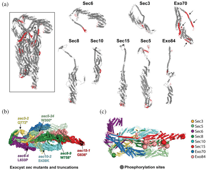
FIGURE 5.

Limited trypsin proteolysis, mutations, and phosphorylation of the assembled exocyst. (a) Limited proteolysis using trypsin identified flexible and exposed regions within the intact exocyst complex. Regions colored in red indicate peptides that were cleaved after an overnight trypsin digest in solution. Arrows point to regions of Exo70 that are buried in the cryo‐EM structure. (b) Regions of exocyst that are truncated or mutated in the original sec mutant screen: 9 sec6‐4 (L633P);11, 50 sec3‐2 is Q772*; sec5‐24 is W300*; sec8‐9 is W758*; sec10‐2 is E439K; sec15‐1 is G836*. Truncated regions are shown as lines. (c) Phospho‐sites mapped to the cryo‐EM structure (Table S3)
