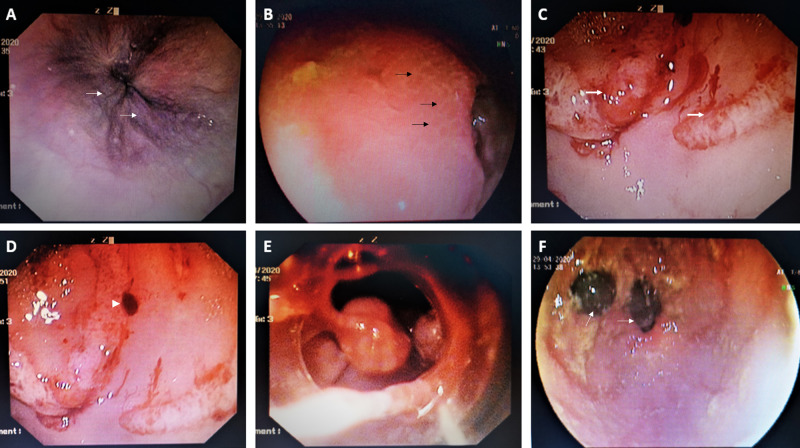
Figure 1. Endoscopic band ligation of actively bleeding portal hypertensive gastropathy.
Urgent upper gastrointestinal (GI) endoscopy showed small low risk esophageal varices without stigmata of recent hemorrhage or active bleeding (A, white arrows) while the gastric mucosa showed extensive mosaic patterning (B, black arrows) of the fundus and body and confluent red spots associated with continuous oozing (C, thick white arrows) and difficult to dislodge fresh clots (D, white arrowhead) suggestive of severe acute bleeding portal hypertensive gastropathy (PHG). Endoscopic band ligation of the bleeding PHG mucosa was performed at targeted points (E) leading to complete hemostasis as seen on relook endoscopy 24 hours later (F, white arrows).

